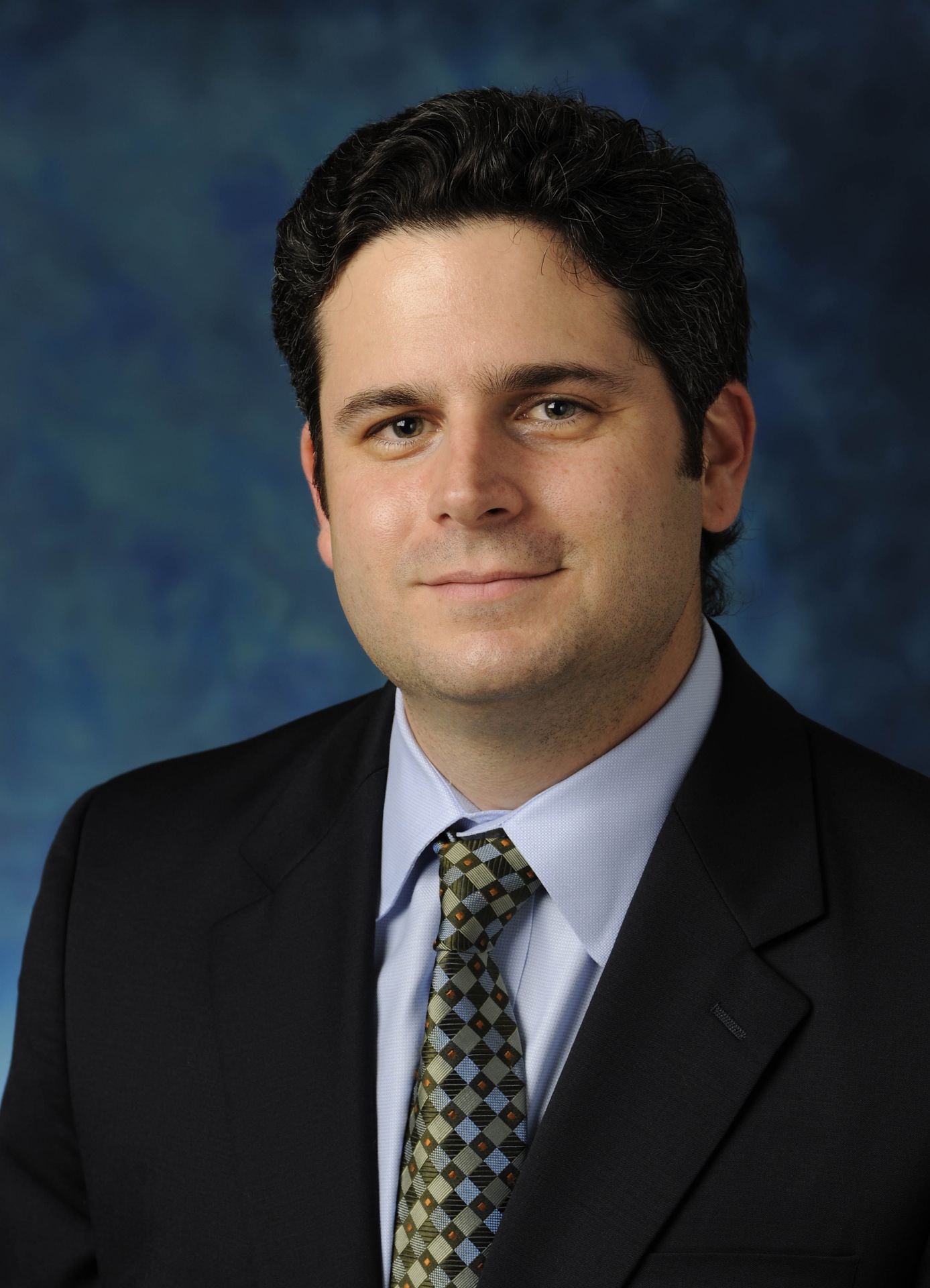
 Michael A. Gleiber, M.D., F.A.A.O.S., is a Board-Certified, Fellowship Trained, Spinal Surgeon and Founder of Michael A. Gleiber, MD, PA, in Jupiter and Boca Raton, Florida. In this article, he is interviewed on his minimally invasive surgical techniques for deformity correction.
 Q: What trends are you seeing in minimally invasive spine surgical techniques today? Is it becoming more common among spine surgeons?
Dr. Michael Gleiber: Minimally invasive spine surgery is becoming "the norm" of how spine procedures can and should be performed for patients. These benefits include safer surgery, less violation of the healthy soft tissues and muscles in the spine, decreased pain requirements, decreased blood loss, more accurate placement of implants and a faster return to work as well as shorter hospital stays.
Minimally invasive technique also has an impact on the hospital global cost analysis, as the patient generally requires a shortened hospital stay and less postoperative pain medication as well as a decreased need for a blood transfusion, which is another reason it has become more common today.
Q: Why did you first incorporate minimally invasive surgery into your practice?
 MG: When I began performing minimally invasive spine surgery, this was initially done through neurosurgical decompressive type procedures. However, having done many of these, I then decided to examine my own results looking at open procedures in the standard fashion that were performed (and still are currently performed by many folks in the spine community) versus my results with minimally invasive spine surgery.
 What I found across the board was that patients that had minimally invasive spine surgery ended up leaving the hospital within a day or two, returning to work earlier and their pain after surgery was significantly decreased compared to patients who have had the same procedure using large open incisions. The infection rate in my practice has always been very low; however it's even lower using minimally invasive spine surgery.
Now, one of the aspects of spine surgery that I am truly passionate about is the advancements made in the field of minimally invasive spine.
Q: What is on the cutting-edge of minimally invasive spine surgical technique today?
MG: Surgeons are beginning to use minimally invasive techniques to perform more complicated procedures, such as deformity and trauma cases. Having trained at Columbia University, New York Orthopaedic Hospital, where I was taught adult degenerative as well as pediatric deformity corrective procedures, I then took this knowledge to my fellowship at the Leatherman Spine Center and built on what I had learned there.
I was always interested in pushing the field toward smaller incisions, better short term and long term outcomes and while we have successfully done this using standard open techniques, what we are seeing more these days is that not only can single- or multi-level lumbar fusions be performed in MIS style with interbody grafts, but also large deformity cases, adult degenerative type, pediatric scoliosis and trauma can be done in the same fashion.
Q: What have you learned about performing minimally invasive techniques for these more complex procedures?
MG: One of the things I've found is that using traditional pedicle screws with our anatomic landmarks requires significant stripping of the soft tissues laterally whereas when we place percutaneous screws, not only are we not violating these soft tissues, but we are also decreasing the chance of potential scar formation that can build up around the neural elements, as well as significantly decreasing blood loss in these larger cases where the deformity curve(s) are of significant magnitude.
After pedicle screws are placed in a percutaneous MIS fashion, most systems now have the option of using the rod, once inserted percutaneously through the screw head tulips, in order to address the deformity. There are times when I will need to address the deformity with anterior releases and/or posterior osteotomies; however, generally speaking, an excellent correction can be performed with rod contouring and levering on the implants themselves without having to do anterior releases or fully stripping the posterior muscles.
Of course this does depend largely on the size and type of the curve as well as the patient's body habitus. The other benefits include the ability to significantly reduce a spondylolisthesis using the reduction systems inherent within the instrument sets. In regards to reduction of the curvature, levering on one rod and using rod benders in situ on one side and then making small changes on the contralateral side allows us to obtain excellent correction of the deformity once the rod has already been placed and the screws preliminarily tightened but not yet locked down. Compression and distraction maneuvers can alleviate the need for in situ rod contouring.
Q: Are there any challenges for performing the minimally invasive techniques for deformity correction?
MG: In terms of long constructs, one of the problems we face while doing this minimally invasive procedure is getting a long construct to fuse and unite at all levels. Therefore, I like using interbody grafts, whether ALIFs or TLIFs, to increase the surface area for fusion in addition to reducing a lateral listhesis or spondylolisthesis, which we can frequently see with adult degenerative deformity.
One thing to note is that decortication of the transverse processes still is very possible when placing your instrumentation percutaneously since the screw tulip is adjacent to the transverse process. Therefore, by palpation and feel of the transverse process, I am able to then decorticate and, if need be, place autograft or allograft on these key structures so that we have a posterolateral fusion in addition to an interbody fusion. In doing this, we have been able to perform surgeries where the blood loss and infection rate has been significantly lessened, and the time on the operating room table is also lessened since all of these screws are placed through radiographic means intra-operatively.
However, it does require a significant commitment, patience and time training to incorporate this into one's surgical armamentarium and have the expected surgical outcomes.
Q: Are there any differences in the preparation work for these cases with the minimally invasive procedures?
MG: When templating these cases, it is highly recommended that patients obtain full thoracic and lumbar X-rays with flexion/extension views, but also lateral bending views to see how mobile the curve is. We should also be aware that many patients who have deformity also have anomalies within their spine anatomy. Therefore, in all my patients preoperatively, in addition to the X-rays, I will order a CT scan of the thoracic and lumbar spine, as well as the sacrum and pelvis to look for pelvic obliquity, vertebral body anomalies and pedicle anomalies, in addition to other congenital defects so that I know when addressing this particular level exactly what how anatomy appears. A three-dimensional CT scan is also extremely helpful in this regard.
Lastly, in regard to neural imaging, it's critically important to have an MRI of the thoracic and lumbar spine to evaluate the spinal cord itself as it may be draped over the vertebral bodies of the apex of the curve and special care needs to be taken when placing screws in these levels.
Q: What do spine surgeons need to know before incorporating these techniques into their practice? How can they work up to that point?
MG: Spine surgeons should be comfortable placing percutaneous screws and reducing grade 1 or 2 spondylolisthesis before considering minimally invasive techniques for deformity correction. One thing I utilized in my practice before embarking on MIS deformity techniques was making sure I was comfortable with one, two or even three level lumbar non-deformity cases with interbody grafts.
Once I became comfortable with this approach over a period of time, I then expanded my surgical practice to include MIS deformity cases and have found it extremely rewarding to obtain the correction and see my patients return to their normal activities of daily living more rapidly than with our standard techniques.
Most importantly, the outcome and your comfort level for deformity cases should never be limited by the size of the incision and if you do require better access, don't hesitate to make the incision larger and convert to an open technique. This is really an important concept in minimally invasive spine surgery.
More Articles on Spine Surgery:
5 Tips for Better Billing in Spine Practices
Running a Spine Practice in the Internet Era: 4 Things to Know
7 Spine Surgeons in New Leadership Roles


