Mastering microsurgical techniques proves essential for spine surgeons performing minimally invasive spinal surgery. The following four spine surgeons provide insight into their acquaintances with the surgical microscope.
Question: How did you start using the microscope for OSS?
K.D. Riew, MD, Columbia Orthopedics, New York City: After I finished my fellowship, I spent two weeks with John McCulloch who taught me how to use the microscope. Initially, I only used it for part of the decompression. As I got more comfortable with it, I used it more frequently and now I use it for everything that I do.
Michael Mayer, MD, PhD, Schön Klinik München Harlaching, München, Germany: I started using the surgical microscope from the beginning of my surgical career. Since I am a board-certified neurosurgeon, as well as an orthopedic surgeon, the surgical microscope has always been an essential tool for spine surgery.
Roger Härtl, MD, New York City Presbyterian Hospital – Weill Cornell Medicine: I'm a neurosurgeon by training, so during our residency, we were exposed to the microscope early on. It became an obvious tool that we would use for cranial surgery and also for spinal surgery.
Mohd Hisam Muhamad Ariffin, MBBS, UKM Specialist Centre, Kuala Lumpur, Malaysia: My first experience with the microscope was actually for hand surgery micro-vascular work as an orthopedic surgeon. It was during a combined ortho and neuro spinal cord tumor surgery; my neurosurgery friend introduced me to using the microscope for spine surgery and from that moment the loupe, which is "the first love," was forgotten.
Q: Why did you start using the microscope for surgery?
MM: The advantages of the surgical microscope are obvious: it provides light and magnification even in deep surgical fields and through small approaches and still enables the surgeon to have a 3D view of the surgical field. If you look through a surgical approach which is smaller than the distance of your naked eyes, you can only have a 2D view, such as with an endoscope.
RH: Because you can visualize especially neuro structures, much better. And you have 3D visualization of the anatomy, with typically excellent illumination and I think it allows you to perform surgery in a limited space much safer and much more efficiently.
MHA: The physical benefit for a surgeon is obvious that I can now operate in a more physiologic posture, meaning that I can maintain my neck and also my lower back in the normal lordosis and this helps reduce the strain on my neck and as the binocular tubes are tiltable and adjustable according to your posture. The ability to stand in more physiological and ergonomic posture has allowed me to do more surgery and more importantly, will also help me grow old protecting my own spine.
The clinical benefit is tremendous and I think the most important, is that it allows me to see very clearly at the depth of the surgical field, especially in minimally invasive spine surgery where the access is very small. The ability to see at the depth is possible as modern surgical microscopes tailored for spine surgery have a longer focal distance along with the coaxial and homogenous illumination, akin like seeing light at the end of a tunnel. The longer optics, and hence a longer working length, is also important as we use long instruments during surgery and we don't want our instruments to keep knocking on our microscope. The surgical microscope also allows better depth perception, which is the main advantage over an endoscope as we are dealing with very delicate structures here and mistakes are not tolerated.
KDR: It gives unparalleled visualization of the cervical field.
Q: What advantages do you see in using a microscope for your surgeries?
a. Patient
RH: The advantages for the patients are that surgeries can be performed with a very maximum focus on the actual pathology. We can magnify the pathology and surrounding anatomical structures and we can really maximize our ability to effectively treat the pathology without affecting surrounding healthy tissues.
MHA: For the patients, the microsurgical technique has allowed them to have a smaller and cosmetically pleasing incision, reduced blood loss and hence, less pain and reduction in analgesic and opioid consumption. They actually have shorter hospital stays nowadays, for example a minimally invasive transformational interbody fusion will only need to stay for one night and daycare surgery for simple microdiscectomy is now a possibility. My patients also actually go back to their activities and work faster, and this improves their well-being and also will take away the fear associated with spine surgery.
KDR: The more thorough a decompression we can do, the better it is for the patient.
MM: Less invasiveness, with less tissue trauma, less blood loss, shorter OR times, less complications, shorter hospitalization and rehabilitation times.
b. Practice
KDR: The better your patients do, the better it is for your practice.
RH: I think it can impact the practice because you eliminate certain downsides of surgery with conventional, open surgery without a microscope. For example, posture-related issues for the surgeon—bending forward, neck pain—occupational disadvantages from standing there for hours in awkward positions. That is all eliminated from using the microscope because you can bring the operative side toward you… you can go through a case in a much more relaxed way, with much less physical effort.
Also, once you have integrated the microscope into your surgical routine, it really facilitates your workflow. You don't struggle with operating room lights and headlights.
MHA: Being in a teaching institution and using the microscope has allowed me to teach my fellows and residents better as the optical system provides symmetrical images for me and the assistant fellow or resident. Incorporating a 3D monitor also allows the rest of the team to actually see the whole surgical procedure, and they have better understanding of the 3D anatomy. Modern microscopes are provided as cameras for good documentation and recording, which is important not only for subsequent teaching and presentation, but also from the medicolegal point of view, as spine surgeries are high-risk surgeries.
c. OR staff
MHA: For the hospital this translates into increased turnover time because of the short OR time, more patients, lesser complications and hence more revenue.
In short these improvements has actually improved safety during surgery tremendously, allows me to reduce the time of surgery and ultimately better outcomes for the patient and will take care of my reputation also.
MM: Bigger learning effect because assistants, as well as the scrub nurse, can follow the MIS surgery on the video screen.
RH: I think there's a huge advantage because you can project the image that you see under the microscope, you can project it on a screen in the OR, and that will help the staff to follow the progress of the surgery and to also allow them to contribute to the surgery, because they can anticipate what the surgery is doing and what the next instrument the surgeon is going to need.
It helps in moving the case along efficiently, because everybody knows exactly what the surgeon is doing. It's a great teaching tool, but it's also a great tool that helps the assistant surgeon to get involved, and for visitors, such as students and other visiting surgeons…they can see in high quality, exactly what I'm doing.
It is a great tool because it allows you to record portions of the surgery, for documentation, analysis and research.
KDR: Everyone in the operating room can see exactly what you are doing. This allows the surgical scrub to learn the procedure, know what the next step is, and to hand you the appropriate instruments.
Q: Which procedures are best suited for use of a microscope?
RH: Any procedure where the area of interest is narrowed down to a few centimeters. A minimally invasive lumbar laminectomy, MIS TLIF procedure…all of these operations are examples of surgeries where the actual target area for the surgery is really a few centimeters, and those are really good examples for things that can be done really efficiently with a microscope. Even if it's a big operation that extends over multiple levels, the microscope can still be useful because at any give time, you only operate in a limited space. In reality, there's no limitation.
MHA: Nowadays, nearly all spine surgeries are done with the microscope, except for the scoliosis surgery. I feel the best procedures for microscope are the anterior and posterior cervical procedures and minimally invasive procedures for low back surgeries.
KDR: While most people think about using the microscope for decompressions, it is also very useful for any procedure that requires better illumination and magnification. Minimally invasive procedures that are done through a small incision are also made easier by the fact that one can have great illumination and the assistant can see exactly what you are doing.
MM: Discectomies (cervical and lumbar); decompression for spinal stenosis, lumbar fusion ALIF, OLIF, transforaminal (TLIF) or PLIF; cervical fusions/disc replacement.
Q: Why are microscopes preferred over Loupes or headlights?
MHA: I think you can't compare the microscope over a loupes and headlight. But the loupe is good at an entrance level into microsurgical world. The main disadvantage is a loupe has a fixed focal distance and a fixed working length. The amount of magnifications a loupe provides falls very short when compared to a microscope. For me in a university hospital, the "sharing of view" is difficult when using a loupe. When my fellows can't see, they can't assist and more importantly they can't learn. And finally with a loupe, I have to consistently flex my neck and this is ergonomically not good for me.
KDR: With Loupes, one has to look down into the wound, which means that at the end of a long operation, the surgeon’s neck can often hurt. With headlights, one has to constantly adjust the beam to get it to line up. In addition, when I used to wear headlights, I would get a headache after a long procedure due to the fact that the strap or the headlight has to be tightened around your skull. In addition, two surgeons with headlights can often hit each other, necessitating a readjustment of the headlight beam. Finally, only one person can look into a small wound with a set of Loupes and headlights.
MM: Loupes are uncomfortable for longer procedures and require additional light sources (e.g. headlamps), which makes it even more uncomfortable. Not convenient for small approaches because of loss of 3D vision.
RH: I would prefer it for the simple reason that the illumination and the image quality is much better with the microscope. I would prefer it because putting on Loupes and headlights and dealing with batteries in the OR, is very cumbersome and usually the quality of the headlights in most ORs is very poor. The amount of magnification you can get with Loupes is very limited. With a microscope, you have better ability to use the microscope to your advantage.
Q: Are there any advantages in terms of building a practice?
MM: If one is not familiar with spinal microsurgery he or she shouldn't build a spine practice.
RH: If patients know that you are trained in microsurgery and you use the latest microsurgical techniques to maximize your ability to perform MIS microsurgery, I think patients with problems related to spinal issues will probably have a preference to seek out a surgeon with those qualifications. It helps you build a robust reputation and practice.
KDR: Using the microscope, I can videotape a procedure as well as take intraoperative photographs. I use these in my talks. Anyone who needs to give talks or do presentations will find value in being able to take intraoperative videos and photographs.
This article is sponsored by Carl Zeiss.
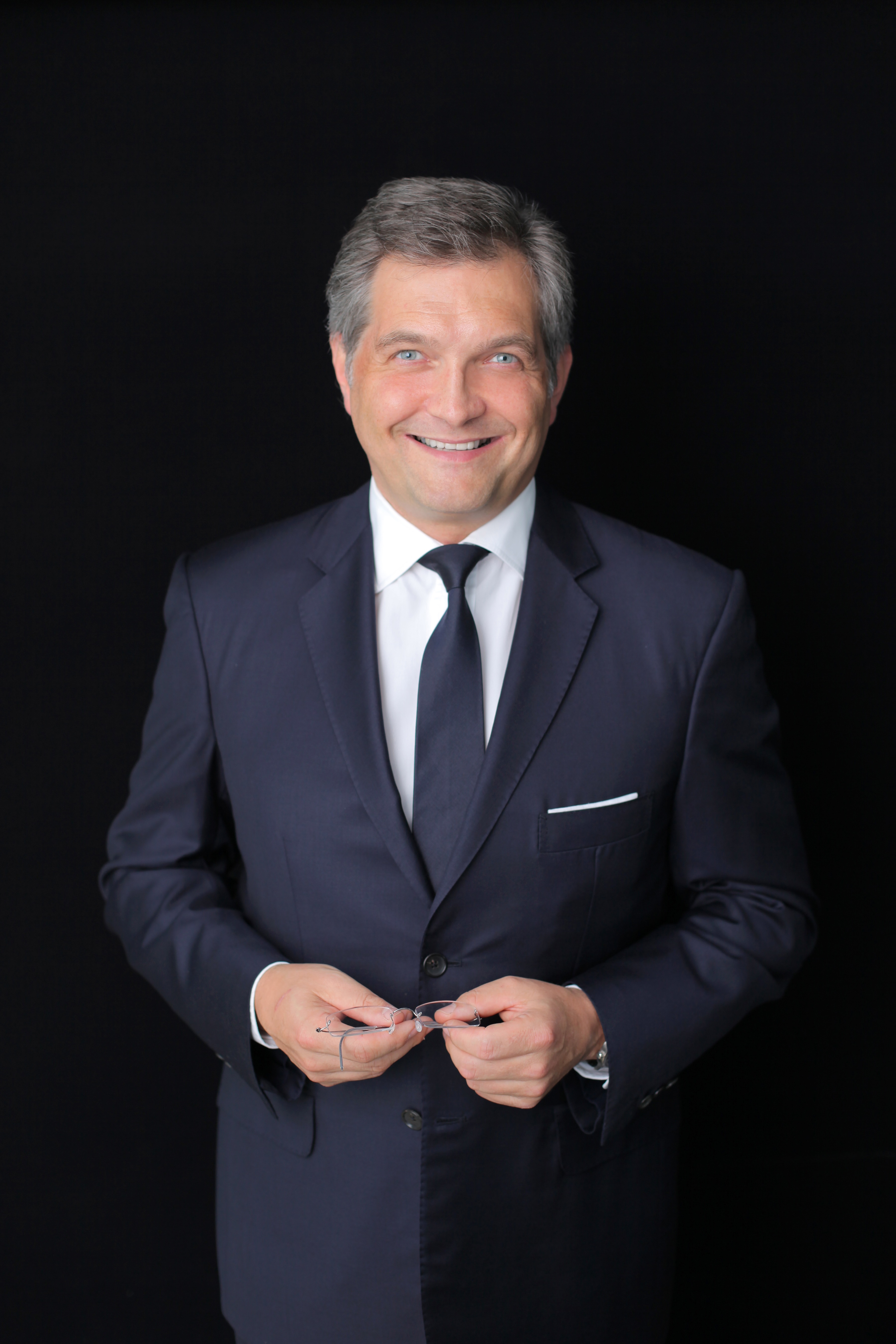
Pictured from left to right: Dr. K.D. Riew, Dr. Michael Mayer, Dr. Roger Härtl & Dr. Mohd Hisam Muhamad Ariffin
.jpg)


