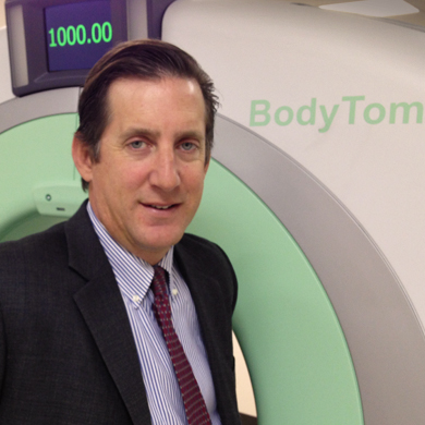
 Eric M. Bailey, MD, is the president, founder and CEO of NeuroLogica, a Boston-based company focused on medical imaging equipment for healthcare facilities and private practices worldwide. He developed the first closed loop micro-machined accelerometer and gyroscope, and holds more than 20 patents for his innovations. Previously, Mr. Bailey was vice president of computed tomography engineering at Analogic Corp., where he developed several medical and security CT systems and was instrumental in developing the first multi-slice CT scanner used in United States airports.
 With NeuroLogica, Mr. Bailey has focused on developing portable CT scanning equipment that can be moved from the imaging room into the operating room, intensive care unit or ambulances to treat patients more efficiently.
"We can pick up patients with strokes and scan them in the ambulances so they get treatment right away," says Mr. Bailey. "We are high quality imaging engineers that are on a real mission in life to change medicine in areas where this type of imaging hasn't been available."
Mr. Bailey discusses how modern imaging technology can improve orthopedic and spine care and where the field is headed in the future.
Q: Orthopedics and spine are rapidly developing fields for all types of technology. How are advancements in imaging equipment making a difference?
Eric Bailey: It's streamlining the surgical process and preventing complications and revision surgeries by bringing the technology into the OR. Our goal is to really give surgeons the gift of three-dimensional X-ray vision; Superman glasses for surgeons. They are super men and women — they have super knowledge, but no one has given them super vision. That's the skills I have in life and our mission is to improve their ability to treat patients.
NeuroLogica has roots in spine, but the equipment can also be used for many other parts of the body. It could have value for knee and complex hip surgery. For example, if there is an older woman who has osteoporosis, this person might fall and break her hip. That hip will fracture like a wine glass and it's like having to put Humpty Dumpty back together again. There hasn't been an X-ray machine that could take a whole picture of the hip at once. Now, with these new developments, CT scanners can get the entire anatomy in one shot.
We're going to find out that this is a very valuable feature going down the road.
Q: How does portable CT scans and three-dimensional technology impact the level of patient care?
EB: These images can help prevent second surgeries and complications. Patients might suffer damage and require more care or compensation for malpractice. Spine and brain surgeons are dealing with a very delicate anatomy and there are some complications. I think this rate will be driven down drastically by having more accurate imaging.
That is also a cost-savings because if you treat the patient better when they are having a stroke or spinal issue and prevent more damage, they get better faster and leave the hospital sooner. This is especially important if the hospital is paid a lump sum for one procedure because if they patients stay at the hospital for five days postoperatively, the hospital loses money. Faster recovery times are more economical for the patient, hospital and insurance economy.
Q: What advantages do surgeons and providers realize from using modern imaging technology?
EB: Surgeons have been using a two-dimensional X-ray for pictures in the past. You only get the picture from one angle and all dimensions are on top of one another. The surgeon has to look at these views and put them together in his or her own brain. With new equipment like the BodyTom, we are able to take large pictures — like the entire spine — that are three-dimensional.
Additionally, the technology is now available to surgeons at any time during the procedure, so they don't have to wait until the surgery is finished to see whether their implants are placed correctly, or whether there is internal bleeding. This is a huge advantage.
Q: How does modern imaging technology change the surgeon's routine and procedure? How can they realize the most clinical benefit?
EB: A lot of patients need a CT scan before surgery, and sometimes they don't realize until surgery has already started that the patient has a life threatening condition. In this case, the patient has to go down for a CT scan in the middle of the procedure while they are still under anesthesia. That can be a life threatening situation, especially considering the delicate area around the spinal cord.
Patients also undergo postoperative CT scans that can show whether the surgeon placed screws or fixation correctly and whether there is any bleeding. They don't know until the images are taken, after the patient has already recovered from anesthesia. The patient is brought down to radiology and that area isn't sterile. Then if the surgeon realizes the screws are placed right or there is a hemorrhage, the patients go back to the operating room.
With emerging technology surgeons can use portable CT scans to take these images in the OR and fix any problems before the patient awakes from surgery. One of the first hospitals where we installed this technology one of the surgeons was doing a complex cervical spine procedure with a four-level fixation. The procedure began and at some point the surgeon took images and saw everything was wrong; they had to start over. The surgery was done properly after that.
If the surgeon didn't catch the initial issue, the patient would have had to do a second surgery. The surgeon said he would never do another spinal surgery case without a CT scanner in the OR.
Q: There is a lot of concern with the cost of care in today's healthcare system. Will purchasing this technology be economically viable?
EB: These developments create an advantage that is cost-effective, and I don't think they will be difficult for hospitals to purchase. One of our first deliveries last year was to the country of Haiti to help communities ravaged by the earthquake. Their conventional CT scanners were no longer capable of running and at any one time, these small battery operated CT scanners were the only operational imaging equipment in Haiti. We shipped them to provide medicine in one of the poorest nations in the world and found a way to make it economically viable there.
Some of our earliest luminaries were also community hospitals in the United States. In hospitals where surgeons perform spinal operations, they tie up the main CT scanner for long periods of time with their patients and others in the waiting room with a non-emergent issue, such as a kidney stone, are left waiting.
Hospitals make a lot of revenue on imaging and it's a very profitable service. With new models available they can perform the CT scans in the OR and bill for them along with the surgeries. It's a real CT scan so they can get paid for those images. There is a revenue stream that can offset the cost of the equipment. To some degree, this is the first step in making these images more affordable.
Finally, spine surgery is also competitive from a marketing standpoint. Four of five people will have back pain that will cause them to have work loss at some point in their lives. A lot of people get to the point where they need medical care and traditionally people would go to their community hospital. Now, they are looking on the internet, TV and other advertisements to find the place that will give them the best care. People move around more and go where they think they'll have superior care.
It's lucrative for hospitals to advertise they have better technology and how their patient outcomes have improved.
Q: Hospitals are facing Medicare rate cuts, and surgeons aren't far behind. Commercial payors often follow government payor trends as well. Will payors support this new technology?
EB: The government and private payors are more supportive of any technology today that can improve the outcomes and shorten length of stay in hospitals. Medicare has been changed under healthcare reform, and they are releasing requirements for hospitals that tie payments to the quality of treatment. Everything is quality-based. They don't want to see negative outcomes, infections or complications because if your numbers are too high they won't receive payment.
Surgeons know medicine is moving to more qualitative indexes that are tied to payment and want to improve their outcomes and patient experience.
Q: Where do you see imaging technology headed in the future?
EB: One of the major innovations in spine surgery was navigation systems. The navigation systems can operate off of preoperative CT images. We have added to that cycle new CT images to make sure the navigation is on track. I perceive a new technology that will add to those two: robotic assisted equipment that will drastically improve the accuracy of the procedure. In urological and gynecological surgery, the da Vinci robot has done that.
There are companies, universities and major researchers investigating robotic equipment to aid in the delivery of screws and needles in spine. That doesn't eliminate the surgeon; it makes them more important and allows them the tools to perform a more accurate procedure. Robots don't have eyes; humans have to see for them and make surgical decisions.
We are adding it all together and I think that's the way things will go in the years to come.
More Articles on Spine Surgery:
5 Steps for Spine Surgeons to Resolve Liability Insurance Before Hospital Employment
5 Policy Steps for Spine Surgeons to Influence Spine Care Policy
9 Mistakes to Avoid When Adding Spine Surgery to an ASC


